
Seeing Clear Through
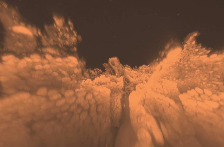
Although the formation seen here could easily pass for a sepia-toned collection of clouds, you won’t be seeing these structures up in the sky anytime soon. The pink wisps are, in fact, fluorescently labeled intestine cells that were imaged within an intact mouse intestine—a feat made possible by a new technique developed by researchers in the lab of Viviana Gradinaru (BS ’05), assistant professor of biology. With this method, researchers can now make thick masses of tissue samples—such as organs and even entire organisms— almost completely see-through, a capability that has numerous research and clinical applications. Rather than having to physically slice through tissue, image each thin slice, and then digitally reconstruct the images into a 3-D visualization of the cells in an organ, researchers using Gradinaru’s technique can bypass these time-consuming steps by applying a solution of detergents to whole organs or organisms. The detergents dissolve light-blocking lipids in the cells, while the structures remain intact thanks to a supporting hydrogel that the researchers embed throughout the tissue—meaning that it becomes possible to look directly through and locate specific cells.